A "GPS" for a Safer, Smarter Way to Repair Your Heart
When you need a procedure to fix your heart or blood vessels, you want the most advanced tools in the hands of leading experts. Some heart procedures, called endovascular procedures, can diagnose or treat your heart disease from the inside. That means we need special tools that allow us to see into your body in real-time to be successful.
Endovascular procedures are often safer than traditional surgery because they only they use small tubes, called catheters, to move around your body and work on targeted areas. The result is less scarring and a quicker recovery.
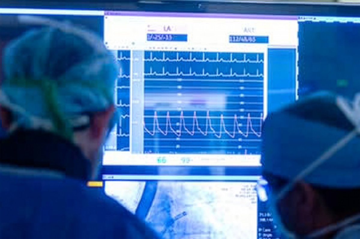
The special tool we depend on to see into your body in real-time is a kind live X-ray called fluoroscopy. But, while helpful, these X-rays expose both patients and the medical team to radiation. “Even though I wear lead and protect myself, I still get irradiated,” notes Behzad Farivar, MD, a heart surgeon and director of UVA Health’s Aortic Center.
Over time, repeated exposure to x-rays is harmful. At UVA Health, we’re tackling this problem by adopting a new technology to replace traditional fluoroscopy for certain procedures. Called Intra-Operative Positioning System (IOPS), this GPS-like tool doesn’t use X-rays to create live images. It’s also more precise than fluoroscopy, which helps us pinpoint the areas needing treatment and get through procedures faster.
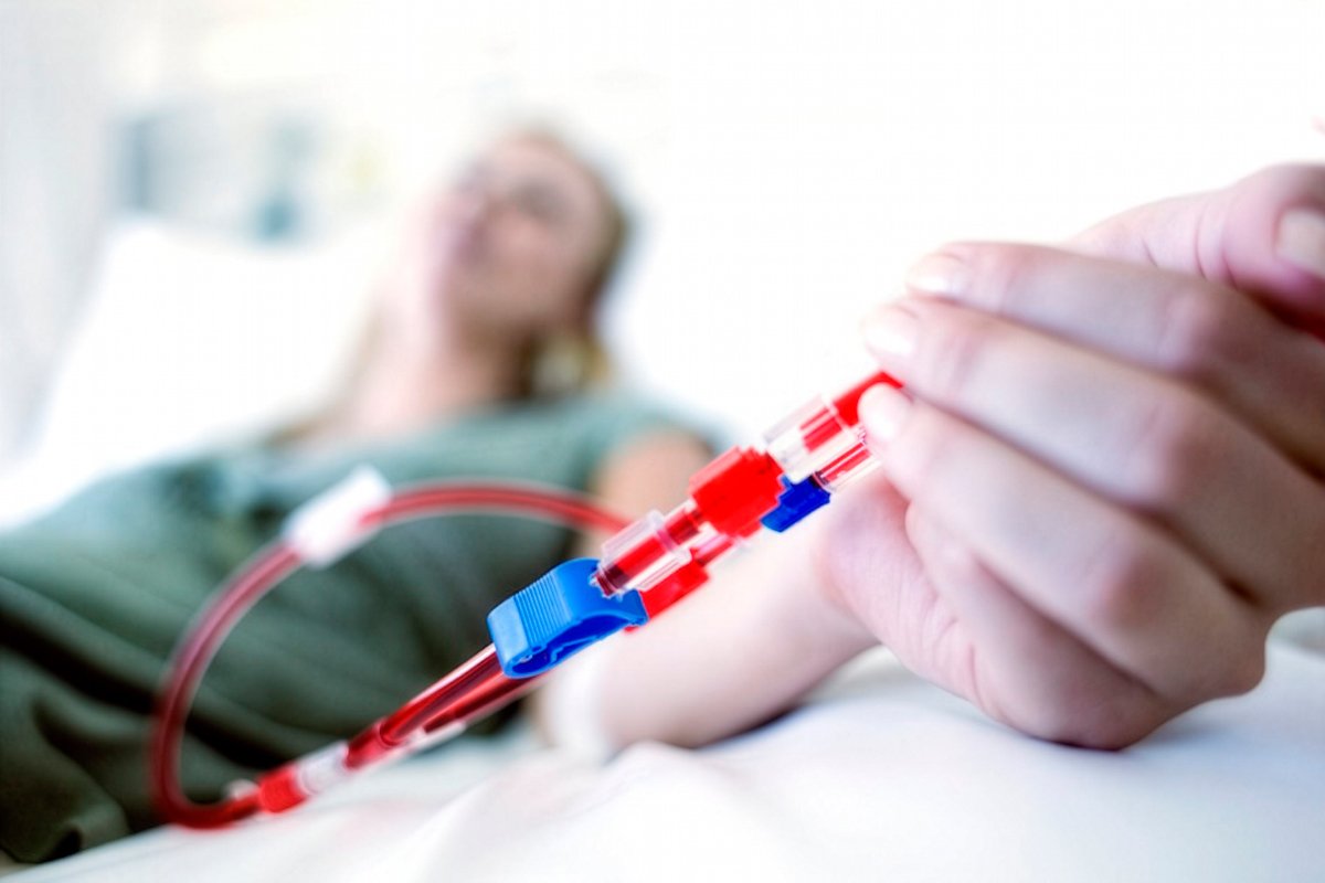
What Is a Catheter?
Besides fluoroscopy, we also rely on catheters to perform endovascular heart procedures. But what are catheters? See more about these important tools.
The Hidden Risks of Fluoroscopy
To avoid large scars, endovascular procedures use your body’s blood vessels as a highway system. In catheters that we put in your blood vessels, we guide cameras and tools to the place in your body where we can treat your heart disease. “But in order for me to do that,” says Farivar, “I need to go from the groin to the aorta and its branch vessels. And that step is all done under fluoroscopy.”
“The longer the procedure, the more complicated it is, and the higher the radiation dose,” he explains. "Over time there's a risk of cancer development with all these radiation exposures." For patients, the risk of cancer can add up if you need multiple imaging tests and treatment procedures over time. And for healthcare providers who are in the operating room every day, this is a serious concern.
A "Win-Win" for Everyone With IOPS
IOPS solves this problem by placing a special tracking pad on the patient, which creates a safe electromagnetic field. This GPS-like system tracks the surgical tools as they move through your body. “It’s an electromagnetic field that is generated, which is non-ionizing radiation,” says Farivar. “So, it's safe both for the patient and the provider.”
Instead of a 2-dimensional (2D), black-and-white X-ray, the surgeon looks at a screen that shows a clear, 3D image of where the catheters are going. With an improved view of the your inside anatomy, we can be more precise and accurate during treatment. “We don't have any limitations and can look at blood vessels from multiple angles. You know exactly where you are inside the body without having to guess, which you don't have that luxury in fluoroscopy,” says Farivar.
While we may still use a small amount of fluoroscopy when needed, IOPS allows us to do most of the procedure without any radiation at all. This creates a safer environment for you and your care team.
Pioneers in Better Heart Care
Farivar performed the very first case using IOPS at UVA Health. He was also was involved in the first ever case in the U.S. that used IOPS to treat a patient. As our techniques and equipment evolve, we’re paving the way for a new era in safer, smarter endovascular heart procedures.