Researcher Highlight Q&A: Jonathan R Lindner, MD, Advanced Heart Imaging & Innovative Therapies
Jonathan R. Lindner, MD, is a physician-scientist researcher who focuses on cardiac imaging and its role in treating cardiovascular disease. Lindner is the current Frances Myers Ball Endowed Professor of Medicine in cardiology and serves as vice chief of research in the cardiovascular division at UVA Health.
Lindner works with a team of experts in vascular biology, noninvasive imaging, microvascular physiology, acoustic physics, and more at his lab in the Robert M. Berne Cardiovascular Research Center. Together, they've helped develop new ways of performing molecular imaging, perfusion imaging, and cardiovascular functional imaging, with the goal of reaching a better understanding of disease pathophysiology and how treatment impacts the disease. Using noninvasive imaging techniques, they've improved patient care through the discovery of new therapeutic targets and the development of new diagnostic algorithms.
See Lindner's selected publications. Below, Lindner discusses his research work and answers our Researcher Highlight questions:
Research Advancements In Cardio Imaging
I absolutely love on a daily basis being able to get into what we call a research sandbox, an area where there's interaction between a diverse set of people with different scientific backgrounds. They include people who are clinicians, people who have expertise in vascular biology, physiology, chemistry, and even acoustic physics.
I'm Jonathan Lindner. I'm a physician scientist in cardiology, and I serve as the vice chief for research in the cardiovascular division. I am a cardiovascular imager.
That is where my research lies in developing new ways of being able to image the heart non-invasively. Many years ago, we developed the field of contrast ultrasound where we use these tiny micro bubbles, smaller than the blood. cells of your body, that ring in an ultrasound field like a musical instrument. These are now used in millions of patients a year to better diagnose heart disease. We're now creating the next generation of ultrasound contrast agents that are smart, that can hone in on disease so we can diagnose it earlier, or even micro bubbles that ring rather violently that we can deliver genes or drugs to specific tissues of the body.
What are you working on right now?
Our lab has 2 major areas of focus. The first is to use advanced forms of imaging, including molecular imaging, to develop innovative therapies for cardiovascular disease. This research involves both preclinical models and human translational studies.
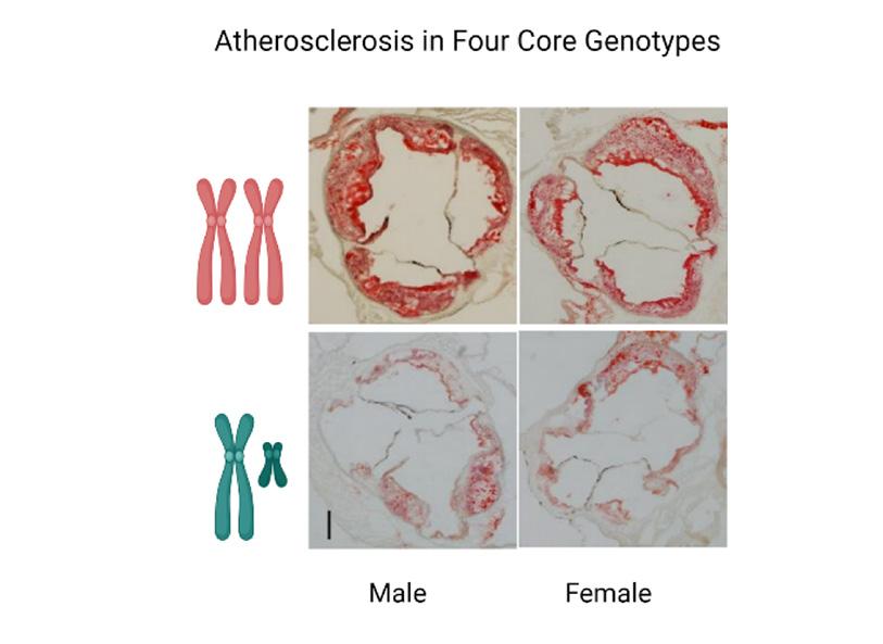
Specifically, we're using advanced imaging to help design therapies to quiesce atherosclerotic disease after acute MI, improve microvascular function, and prevent the progression of aortic stenosis.
Our lab’s other major focus is to develop new therapies based on ultrasound cavitation (collapse of microbubble contrast agents) to either augment perfusion in ischemic tissues or enhance site-specific delivery of vectors for gene therapy.
What are the most intriguing potential clinical applications of your work?
We believe that the use of advanced imaging as a research tool, particularly for the evaluation of thromboinflammatory processes, will lead to unique methods for preventing remote plaque activation after an ischemic event, and for preventing the progression of aortic stenosis once it is diagnosed at an early stage.
Our ability to noninvasively assess microvascular physiology and molecular phenotype is starting to yield important information vital for the implementation of precision medicine in microvascular dysfunction and cardio-oncology, and even to mitigate the effects of space radiation in astronauts on long-duration space missions.
With respect to ultrasound therapeutics, we are now actively translating studies using cavitation to augment limb perfusion in patients with peripheral artery disease and critical limb ischemia. We're also poised to begin translation of using cavitation to enhance cardiomyocyte transduction with adeno-associated virus vectors designed to treat certain forms of cardiomyopathy.
What made you choose UVA Health as the place to do your research?
I recently returned to UVA, where I started my career, for 2 major reasons. First, here I am surrounded by so many talented, innovative, and bold basic scientists and physician-scientists in the fields of vascular biology, cardiovascular physiology, and cardiovascular imaging. More importantly, these same individuals are the most kind, supportive, collaborative, and happy people you will ever meet in academic medicine.
What do you wish more people knew about your area of research?
I wish more people realized how advances in noninvasive imaging that are intended for diagnostic applications in patients can also be leveraged to accelerate knowledge of disease pathobiology and treatment. Advanced imaging can be an incredibly impactful research accelerator, which is vital to improving the efficiency of and lowering costs for drug development.
How did you become interested in your area of research?
By working with several mentors with diverse backgrounds in noninvasive imaging, vascular physiology, and vascular biology. I gradually came to realize that, as a physician-scientist, putting together these 3 disciplines could lead to some unique understandings of disease and therapy.
Subscribe to Healthy Practice
Get UVA Health news & information specifically for referring physicians